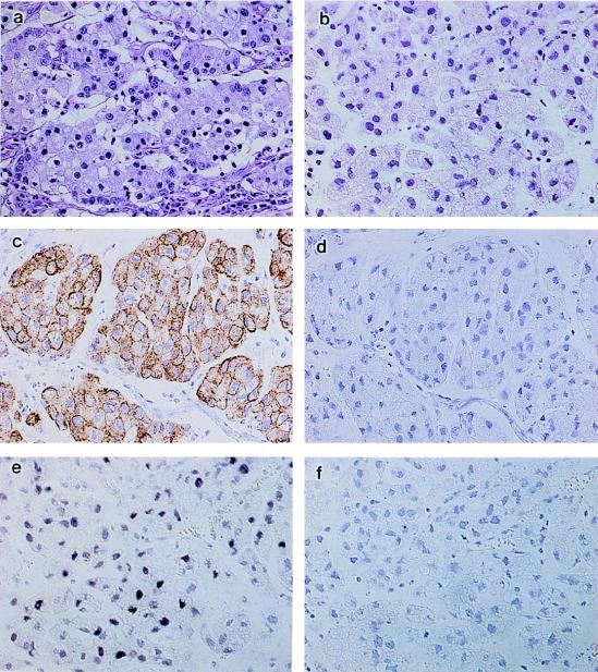
FIG. 1.
Morphological evaluation of a human EBV-associated gastric carcinoma (EBVaGC) transplanted to SCID mice. (a) The original EBVaGC is a poorly differentiated adenocarcinoma with solid nests and lymphoid infiltration. (b) The transplanted tumor in SCID mice, designated the KT tumor, lacks lymphoid infiltration, and half of the tumor consists of mucus-producing cells, which were a minor component of the original tumor. Like the original tumor, the KT tumor shows cytokeratin immunoreactivity in the cytoplasm (c) but no positive staining when the cytokeratin antibody was replaced with mouse immunoglobulin (d). The KT tumor also shows a positive signal in the nuclei with EBER1 ISH (e) but no signal when the antisense oligoprobe was replaced with a sense probe (f). Magnification, ×80.

