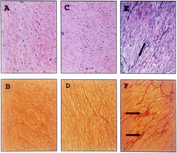
FIG. 5.
Histology of myocardial tissue from pCMV/VP1- and VV-VP1-vaccinated mice after CVB3 challenge. Paraffin sections from heart tissue of pCMV/VP1 (A and B)- and VV-VP1 (C and D)-vaccinated and nonimmunized (E and F) BALB/c mice 45 days following challenge with 5 LD50 of CVB3 for the immunized mice and 1 LD50 of CVB3 for the nonimmunized mice were stained with hematoxylin-eosin (A, C, and E) or Sirius red (B, D, and F); connective tissue is stained red. No infiltrating immune cells or unusual high levels of connective tissue, indicating ongoing myocarditis or fibrosis, were detectable in immunized mice. In contrast, fibrotic tissue, indicating former virus-caused tissue destruction, was present in heart samples from nonimmunized mice (E and F, arrows). Magnification, ×125.

