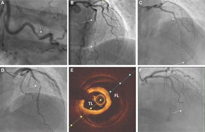
Figure 3.
Angiographic appearances of spontaneous coronary artery dissection: (A) Type 1—characterized by dual lumen appearance and contrast hang-up due to contrast penetration of the false lumen. (B) Type 2A—long narrowing (arrows) often tapering at the distal extent and with recrudescence of a normal calibre vessel distally. (C) Type 2B—long narrowing (arrows) which extends distally with- out restoration of a normal calibre vessel distally. (D) Type 3—shorter stenosis (arrow) angiographically difficult to distinguish from focal atherosclerotic plaque without confirmation by intra-coronary imaging. (E) Optical coherence tomography (OCT) image from case shown in (D). TL, true lumen; FL, false lumen. (F) Type 4—Occlusion, often distally. May be difficult to distinguish from coronary embolus but may have upstream tapering to suggest an underlying occlusive intramural haematoma.

