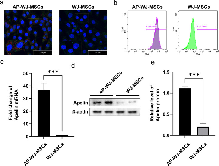
Fig. 1.
Overexpression of apelin in WJ-MSCs and analysis of transduction efficiency. (a) Immunofluorescence images showing the intracellular GFP fluorescence in AP-WJ-MSCs (Blue: nuclear DAPI, Red: Apelin, Green: GFP). (b) Quantitative flow cytometric analysis confirming the efficiency of lentiviral transduction in WJ-MSCs. (c) qRT-PCR results indicating a 36.74-fold increase in Apelin gene expression in Ap-MSC-sEVs compared to that in MSC-sEVs (***P < 0.001). (d–e) Western blot images depicting the protein expression levels of apelin in AP-MSC-sEVs (***P < 0.001). Abbreviations: sEVs, small extracellular vesicles; AP-MSC-sEVs, apelin-MSC-sEVs, engineered sEVs loaded with overexpressed apelin; Wharton’s jelly-derived mesenchymal stem cells (WJ-MSCs)

