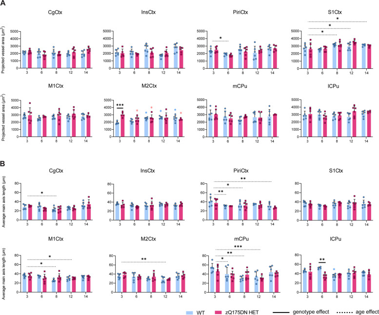
Fig. 8.
Blood vessel area and vessel length in the zQ175DN HET mice. (A) Projected vessel area (μm2) relative to FOV and (B) average main axis length (μm) in WT and zQ175DN HET mice at 3, 6, 8, 12 and 14 months of age for 8 brain regions, represented with subject values and a group mean ± SD (two-way ANOVA, p < 0.05); Colored asterisks represent the overall age effect within WT (blue) and zQ175DN HET (red), compared to 3 months. Significant difference after FDR correction * p ≤ 0.05, ** p ≤ 0.01, *** p ≤ 0.001, **** p ≤ 0.0001; CgCtx—Cingulate Cortex, InsCtx – Insular Cortex, Piri – Piriform Cortex, M1/2Ctx – Motor Cortex 1/2, S1Ctx – Somatosensory Cortex 1, CPu – Caudate Putamen, medial (mCPu) and lateral portion (lCPu)

