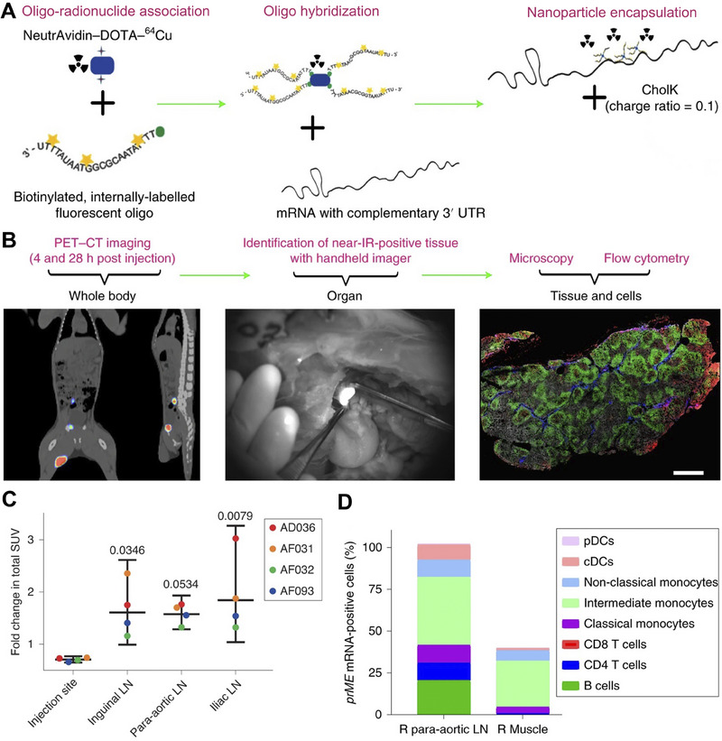
FIGURE 14.

Whole‐body PET‐CT imaging and fluorescent labeling method of mRNA delivery in vivo. A Labeling mRNA with dual radionuclide‐near‐infrared probe. B Whole‐body PET‐CT imaging, mRNA‐positive tissues near‐infrared identification, and protein expression analysis after administration. C Fold change in total SUVs over 28 h in different sites of 4 cynomolgus macaques (AD036, AF031, AF032, AF093). D DCs and B cells accounted for the predominant labelled‐mRNA‐positive cell types. Reproduced with permission.[ 196 ] Copyright 2019, Nature Publishing Group.
