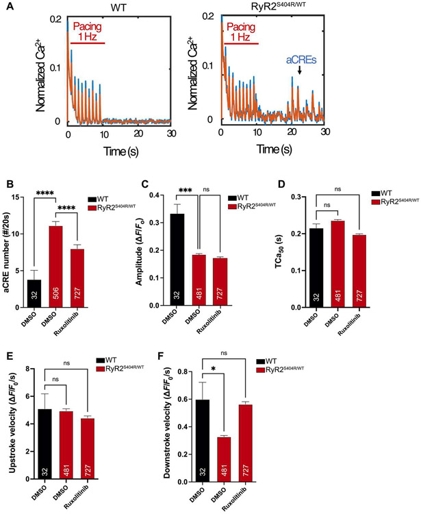
Fig. 5. Ruxolitinib suppresses abnormal calcium signaling in a cellular model of CPVT.
iPSCs with the dominant RYR2 mutation (p.S404R) were generated from a patient with a clinical diagnosis of CPVT (RYR2S404R/WT) and differentiated into functional CMs (iPSC-CMs). After plating on glass-bottom dishes, RYR2S404R/WT and WT iPSC-CMs were loaded with the fluorescent Ca2+ indicator (Fluo-4) and electrically paced at 1 Hz for 10 s. (A) Representative tracings of calcium transients in WT and RYR2S404R/WT cells. The appearance of aCREs after the cessation of pacing is indicative of deranged Ca2+ signaling in CPVT iPSC-CMs (right). Raw data are in blue, and filtered data are in orange overlay. (B) Quantification of aCREs over 20 s in WT and RYR2S404R/WT iPSC-CMs with preincubation of 2 μM ruxolitinib compared with vehicle alone (DMSO). (C to F) Automated analysis of Ca2+ transient parameters during 1-Hz pacing of iPSC-CMs for mean amplitude (C), Ca2+ transient duration at 50% of peak amplitude (TCa50) (D), upstroke velocity (E), and downstroke velocity (F). The numbers of analyzed cells (n) are annotated on each graph and were from at least two independent differentiations and four separate culture wells. Statistics were performed by one-way Kruskal-Wallis with Dunn’s multiple comparisons test: ns, P > 0.1; *P < 0.05; **P < 0.01; ***P < 0.005; and ****P < 0.0001.

