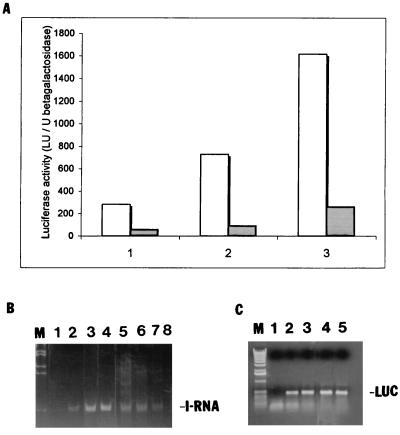
FIG. 5.
HCV-luciferase reporter dose response and quantitation of IRNA and luciferase mRNA in the cell line. (A) Increasing concentrations (1, 2, and 3 μg) of the pCD HCV-luc reporter plasmid were transfected into either Huh-7 hepatoma cells (dotted bars) or the IRNA-expressing hepatoma cell line (pCDIR.Ribo.ΔT7) (white bars). A control β-Gal plasmid was also cotransfected to normalize transfection efficiencies. Luciferase activity (103 light units [LU]) was plotted against increasing concentrations of the test plasmids. (B) Detection of IRNA in hepatoma cell line by RT-PCR. IRNA expression level was detected by RT-PCR using IRNA-specific oligonucleotide primers. Lane 1, no-RNA control; lanes 2 to 4, 1, 2.5, and 5 ng, respectively, of in vitro-transcribed, purified IRNA; lanes 5 to 7, 2, 1.5, and 1 μg, respectively, of total RNA from an IRNA-expressing hepatoma cell, pCDIR.Ribo.ΔT7. Lane 8 contained 2 μg of total RNA from the control Huh-7 cells. Lane M represents marker DNA. (C) Detection of luciferase (LUC) mRNA by RT-PCR. Total RNA isolated from control (Huh-7) and pCDIR.Ribo.ΔT7 cells transfected with the luciferase reporter plasmid was used to detect luciferase mRNA levels by using luciferase-specific oligonucleotide primers. Lane 1, no-RNA control; lanes 2 and 3, 1 and 10 ng of luciferase mRNA standard; lanes 4 and 5, 1 μg of total RNA isolated from Huh-7 cells and cells expressing IRNA, respectively, after transfection with pCDHCV-luc.

