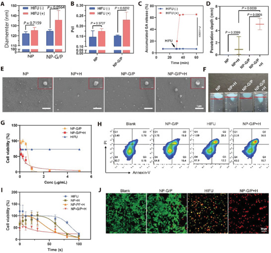
Figure 3.

A,B) The size (A) and polydisperse index (PdI) (B) of NP‐G/P under HIFU irradiation (n = 3). C) On‐demand GA release from NP‐G/P induced by HIFU treatment (n = 3). D) The penetration depth of NP‐G/P after HIFU irradiation in the stimulated tumor extracellular matrix (n = 3). E) Scanning electron microscope (SEM) confirmed the morphological change of HIFU‐treated NP‐G/P. The red box referred specifically to changes in the structure of individual particles. F) The propulsion trajectory of NP‐G/P and NP was measured in the stimulated tumor extracellular matrix. G) The effect of HIFU on in vitro cytotoxicity of NP‐G/P was evaluated by the CCK‐8 method (n = 3). H) Flow cytometry analysis of tumor cell apoptosis after different treatments. I) The effect of nanomotors on the working efficiency of HIFU therapy was evaluated by the CCK‐8 method (n = 3). J) Representative fluorescence images of live (green) and dead (red) 4T1 cells after different treatments. In all cases, significance was defined as P ≤ 0.05. The significance of the difference between two groups was determined via Student's t‐test. The significance of the difference of more than two groups was determined via One‐way ANOVA test.
