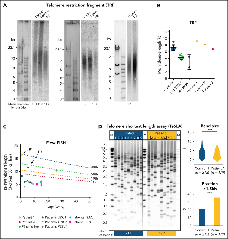
Figure 1.
Telomere length in blood cells from patients. (A) Telomere length determined by telomere restriction fragment (TRF) assay in DNA from blood cells from patients P1, P2, P3 and their parents (P3′s father's sample was not available). (B) Graphic representation of TRF data obtained in panel A as well as in 8 age-matched controls, 8 RTEL1-deficient patients, and 2 PARN-deficient patients described elsewhere.31, 32, 33, 34 (C) Relative telomere length in comparison with the tetraploid control cell line (1301) measured by Flow-FISH in blood cells from P1, P3, P3′s mother and 13 DC/HH patients with pathogenic variants in DKC1, TERT, TERC, RTEL1, or TINF2. Lines represent the 1st, 10th, 50th and 90th percentiles of telomere length of healthy controls. (D) (Left) Detection of the shortest telomeres by TeSLA performed in blood cells from P1 and an age-matched healthy donor. (Right) Graphic representation of TeSLA data with statistical analyses reveals a significant increase in telomere loss events in P1's blood sample. A 2-tailed Student t-test was used for statistical analyses of band size and a χ2 test was used for analysis of fraction < 1.5kb.

