Figure 1.
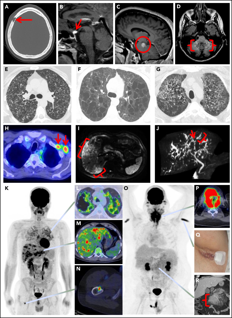
Spectrum of manifestations of LCH in adults. (A) Axial computed tomography (CT) of the calvarium demonstrating a lytic lesion with a beveled edge (arrow). (B) Sagittal pituitary T2 weighted fluid-attenuated inversion recovery (FLAIR) magnetic resonance imaging (MRI) highlighting a hypothalamic lesion with increased FLAIR signal (arrow). (C) Sagittal T1 weighted contrast-enhanced brain MRI demonstrating an enhancing posterior midbrain/pons lesion (circle). (D) Axial brain MRI with a neurodegenerative pattern of T2 signal abnormality throughout the bilateral cerebellar peduncles (brackets). (E-G) Axial CT of the mid to upper chest demonstrating variable appearances of pulmonary LCH to include the most common nodulocystic pattern ground-glass nodules and cysts (E), a cystic-predominant pattern with larger irregular cysts and a few scattered nodules (F), and more confluent combined cystic and nodular disease with architectural distortion consistent with elements of fibrosis (G). (H) Axial fused 18F-fluorodeoxyglucose positron emission tomography CT (FDG PET/CT) of the chest demonstrating 2 FDG avid left axillary lymph nodes (arrows). (I) Diffusion-weighted MRI of the liver demonstrating an infiltrative pattern of increased signal throughout the right hepatic lobe (brackets). (J) Magnetic resonance cholangiopancreatography demonstrating multifocal stricturing (arrow) and dilatation (bracket) of the intrahepatic ducts. (K) Maximum intensity projection FDG PET/CT demonstrating FDG avid advanced pulmonary disease (L), multifocal FDG avid hepatic disease (M), and an FDG avid lytic right femur lesion (N). (O) MIP FDG PET/CT demonstrating an infiltrative FDG avid laryngeal mass (P), bilateral axillary dermal lesions with associated left axillary photograph (Q), and a mildly FDG avid mesenteric mass highlighted with a bracket (R).

