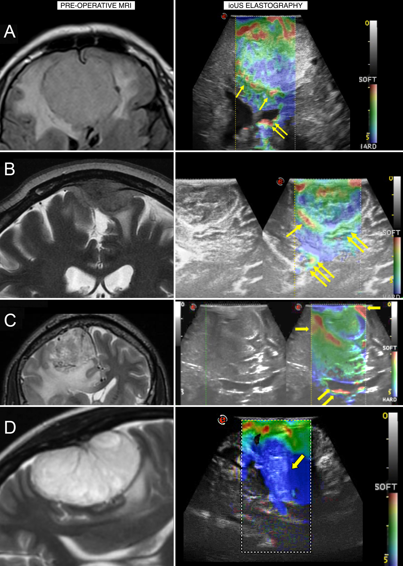
Figure 3.

A) An ethmoidal meningioma displaying a heterogeneous consistency on elastography, suggesting that debulking should begin from softer areas. B) A parietal meningioma with a mixed brain–tumor interface, suggesting that dissection should progress from the better-preserved interface to the most adherent interface. C) A parasagittal meningioma where the elastography assessment of interface conflicts with the preoperative MRI and illustrates a well-preserved interface to begin debulking. D) A convexity meningioma shown on elastography to be harder than visualized on MRI, allowing the surgeon to change from a piecemeal to en bloc resection. ioUS, intraoperative ultrasound. Reprinted from Pepa GM Della, Menna G, Stifano V, et al. Predicting meningioma consistency and brain-meningioma interface with intraoperative strain ultrasound elastography: a novel application to guide surgical strategy. Neurosurg Focus. 2021;50(1):1–11.[40] Permission will be obtained at next stage of manuscript.
