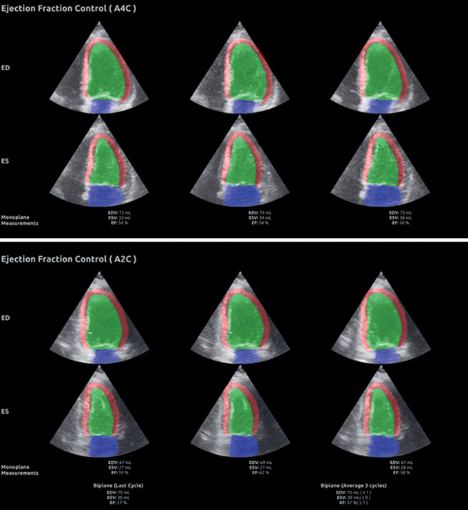
Figure 2.
Summary screen of the segmentation masks with LV volumes and EF measurements. The upper panel shows the segmentation masks with measurements of LV volumes at end-diastole and end-systole as well as EF for three consecutive cardiac cycles obtained from apical four-chamber view. The lower panel shows the similar results for apical two-chamber view. At the bottom of the lower panel, the AI measurements based on biplane recordings are shown.

