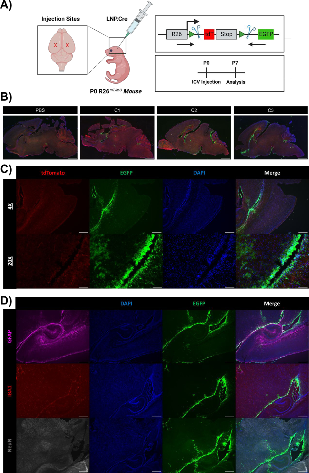
Fig. 2 |. Cellular tropism of C3 LNPs in the neonatal mouse brain.

(A) Scheme demonstrating application of LNPs encapsulating Cre mRNA to P0 R26mT/mG neonates via bilateral ICV injection. Genome modulation in the R26mT/mG mouse model and experimental scheme are also visualized in the right panel. (B) Whole brain histology of neonatal brains 7 days after PBS, C1, C2, or C3 LNP ICV injection, displaying unedited (tdTomato+) and edited (GFP+) regions, imaged at 1X. (C) Histology focused on the brain ventricular lining 7 days after C3 LNP ICV injection, imaged at 4X and 20X. (D) Histology focused on the brain ventricular lining with intracellular staining to capture successful C3 LNP-mediated delivery (GFP+) to astrocytes (GFAP+), microglia (IBA1+), and neurons (NeuN+). Scale bars: 1 mm (1X), 200 μm (4x) and 50 μm (20x).
