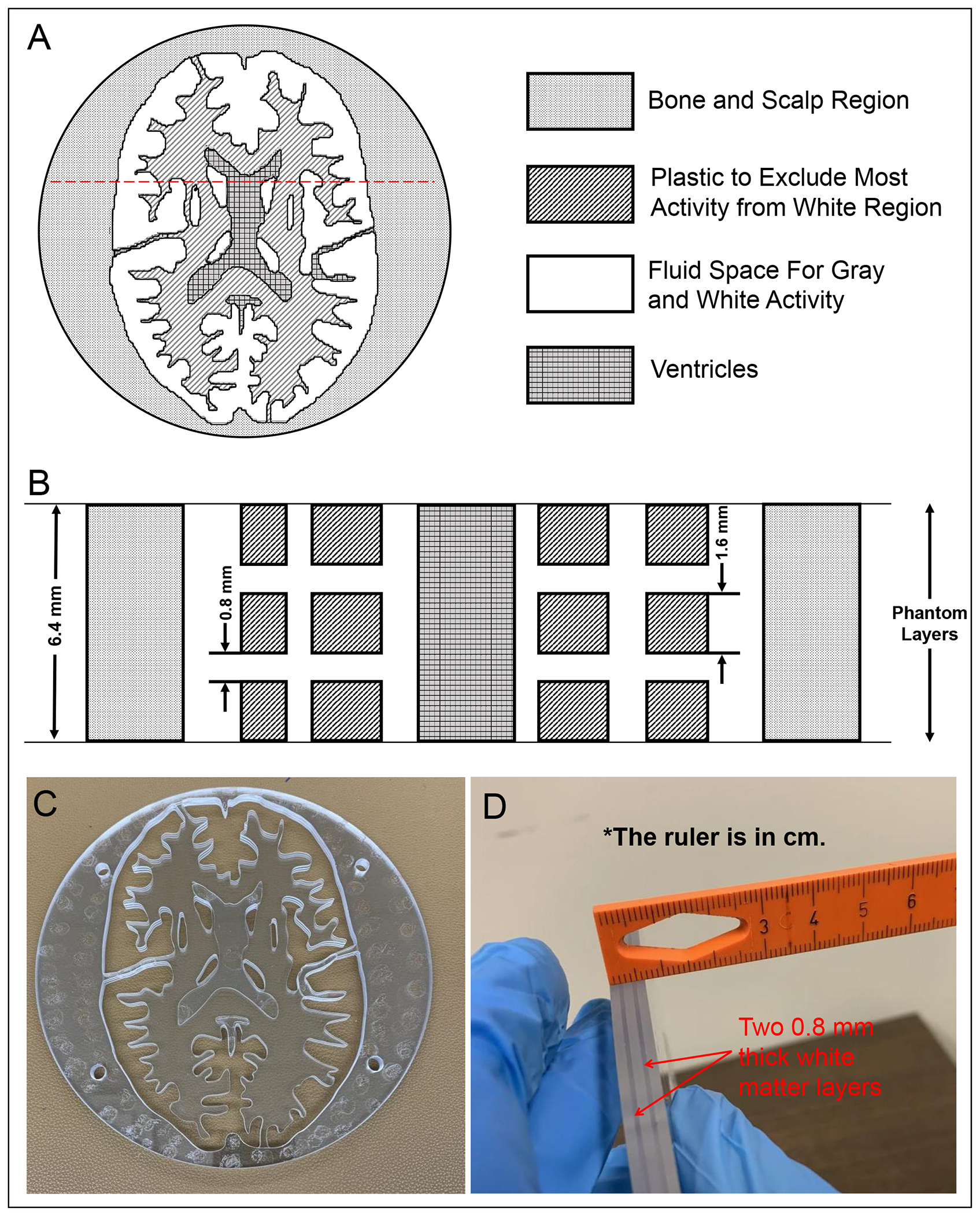
Figure 2.

(A) One representative axial section through the 3D Hoffman brain phantom’s DRO containing two fluid regions (i.e., gray matter and white matter), which will be filled with 18F activity, and ventricles. (B) Schematic coronal cross section through the red dashed lines in A. Gray matter and ventricles extend across the entire 6.4 mm thickness of each slice whereas the two white matter layers are each 0.8 mm thick. Photographs of the representative slice in the (C) axial and (D) coronal views.
