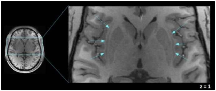
Fig 1. An axial view of the human claustrum.

The human claustra are shown (signaled by blue arrows) between the external and extreme capsule, left and right claustra in a T1-weighted image of one of the participants are shown in MNI coordinates. This figure was acquired using Fslview, from the FMRIB software Library (FSL) tools v5.0 [48].
