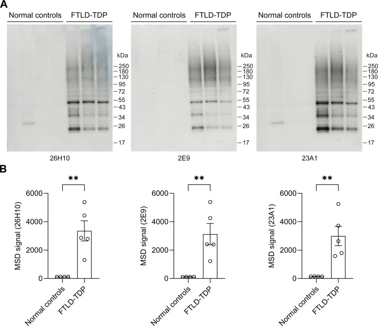
Fig 4. Rabbit mAbs against pS409/410-TDP-43 detect the pathological accumulation of phosphorylated TDP-43 in FTLD-TDP brain tissues.
(A) Immunoblot analysis of urea-soluble fractions from the frontal cortex of FTLD-TDP patients and normal controls using the indicated rabbit mAbs. (B) MSD analysis of phosphorylated TDP-43 protein levels in urea-soluble fractions from the frontal cortex of FTLD-TDP patients and normal controls using the indicated rabbit mAbs (n = 4−5 per group). Data shown as the mean ± SEM. ** (left to right) P = 0.0050, 0.0091 and 0.0075, unpaired two-tailed t-test.

