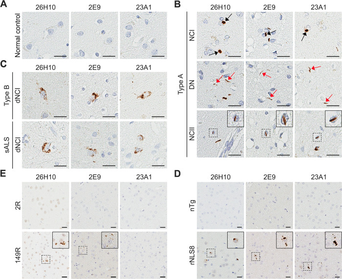
Fig 5. Rabbit mAbs against pS409/410-TDP-43 detect TDP-43 pathology in brain tissues from FTLD and ALS patients and rNLS8 mice.
(A‒C) Representative images of immunohistochemical analysis using the indicated rabbit mAbs against pS409/410-TDP-43 in the frontal cortex of normal controls (A), FTLD-TDP type A patients (B), and FTLD-TDP type B patients (C), and in the motor cortex of ALS patients (C). Black arrows indicate neuronal cytoplasmic inclusions (NCI), and red arrows mark dystrophic neurites (DN). Inserts in B are higher magnifications of neuronal intranuclear inclusions (NCII). (D) Representative images of immunohistochemical analysis using the indicated rabbit mAbs against pS409/410-TDP-43 in the cortex of non-transgenic (nTg) and rNLS8 mice. Inserts are higher magnifications of NCI. (E) Representative images of immunohistochemical analysis using the indicated rabbit mAbs against pS409/410-TDP-43 in the cortex of AAV-2R and AAV-149R mice. Inserts are higher magnifications of inclusions. For all panels, scale bars are 20 μm.

