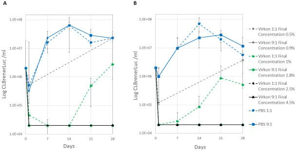
Fig 2. Virkon inactivation of T. cruzi epimastigotes in blood (A) and RTH media (B).
T. cruzi CLBrener Luc epimastigotes were incubated for 1 h with 1, 2 and 5% Virkon 1:1 (dashed lines) and 9:1 (solid lines) volumetric excess giving a final Virkon concentration range of 0.5–4.5%. Cultures were monitored for parasite growth over 28 days (three technical replicates, mean ± standard deviation, limit of quantitation 2e4 ml-1). PBS controls were included.

