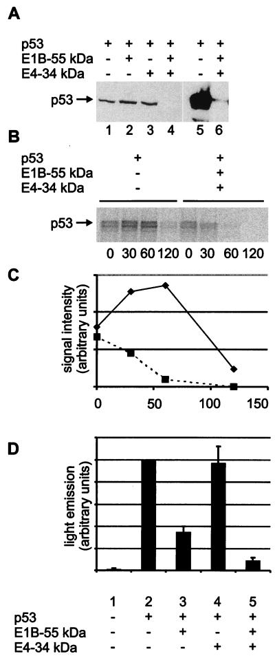
FIG. 1.
Degradation of p53 by the adenovirus type 5 E1B 55-kDa and E4 34-kDa proteins. (A) Expression plasmids for the p53 (0.5 μg), E1B 55-kDa (0.5 μg), and E4 34-kDa (1.0 μg) proteins or “empty” vector constructs were transfected into Saos-2 cells as indicated. After 24 h, the cells were lysed and subjected to SDS-PAGE and Western blot analysis. p53 was detected with monoclonal antibody Pab1801 (lanes 1 to 6). In a second experiment (lanes 5 and 6), the film was overexposed to allow detection of residual p53 in the presence of the E1B 34-kDa and E4 34-kDa proteins. (B) Expression plasmids for the p53 (1 μg), E1B 55-kDa (330 ng), and E4 34-kDa (660 ng) proteins or “empty” vector constructs were transfected into Saos-2 cells as indicated. After 24 h, the cells were labeled with [35S]methionine and [35S]cysteine for 10 min and then incubated in nonradioactive medium (chase). After the time points indicated (minutes), the cells were harvested and subjected to immunoprecipitation with monoclonal antibody Pab421 directed against p53, followed by SDS-PAGE and autoradiography. Note that the signal intensities obtained with p53 alone initially increase, possibly reflecting the incorporation of radioactively labeled amino acids that were internalized into the cells but not yet assembled into protein at the start of the chase. (C) The signal intensities obtained in the same experiment with p53 in the absence (diamonds) or presence (squares) of the E1B 55-kDa and E4 34-kDa proteins were quantified with a Bio Imaging Analyzer (Fuji) and plotted against the time after removal of the radioactive medium. (D) Expression plasmids for the p53 (50 ng), E1B 55-kDa (1.0 μg), and E4 34-kDa (0.5 μg) proteins were transfected as indicated into Saos-2 cells along with a reporter plasmid containing a p53-responsive promoter driving luciferase expression (pBP100luc [27], 0.5 μg). After 24 h, the cells were lysed and subjected to a luciferase assay. Luciferase activity is indicated in relative units, and the value obtained with p53 in the absence of antagonists was set to 100%. Error bars reflect the standard deviation of at least three independent experiments.

