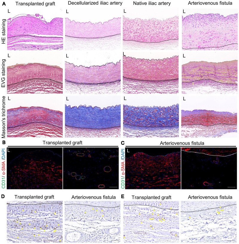
Figure 1.
Evaluation of allogeneic decellularized graft in the human body 5 years post-implantation. (A) Hematoxylin and eosin (HE), Verhoeff’s Van Gieson (EVG) staining and Masson’s trichrome staining of transplanted graft, decellularized iliac artery, native iliac artery and arteriovenous fistula (AVF) (scale bar, 100 μm). (B) Immunofluorescence was used to assess the expression of CD31 and α-SMA in the intima-media (left) and adventitia (right) of transplanted graft (scale bar, 100 μm). (C) Immunofluorescence was used to assess the expression of CD31 and α-SMA in the intima-media (left) and adventitia (right) of AVF (scale bar, 100 μm). (D) Immunohistochemistry (IHC) was used to assess the expression of CD68 in the adventitia of both the transplanted graft and AVF (scale bar, 100 μm). (E) IHC was used to assess the expression of CD3 in the adventitia of both the transplanted and AVF (scale bar, 100 μm). L represents the lumen, the area outside the dashed lines represents adventitia area of native vascular or transplanted graft, and the yellow triangle represents cells with positive expression.

