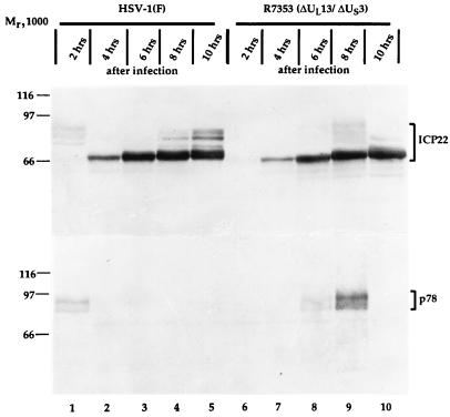
FIG. 7.
Photographic image of cell extracts infected with wild-type virus or with recombinant R7353, separated in denaturing gels and reacted with sera to ICP22 and p78. Synchronized HeLa cells were infected at 0.5 h after release from block with either HSV-1(F) (lanes 1 to 5) or recombinant R7353 (lanes 6 to 10), and extracts were prepared at the times indicated after infection. Cell extracts were separated in duplicate 7% denaturing polyacrylamide gels, transferred to nitrocellulose sheets, and reacted with sera to ICP22 (upper panel) and p78 (lower panel).

