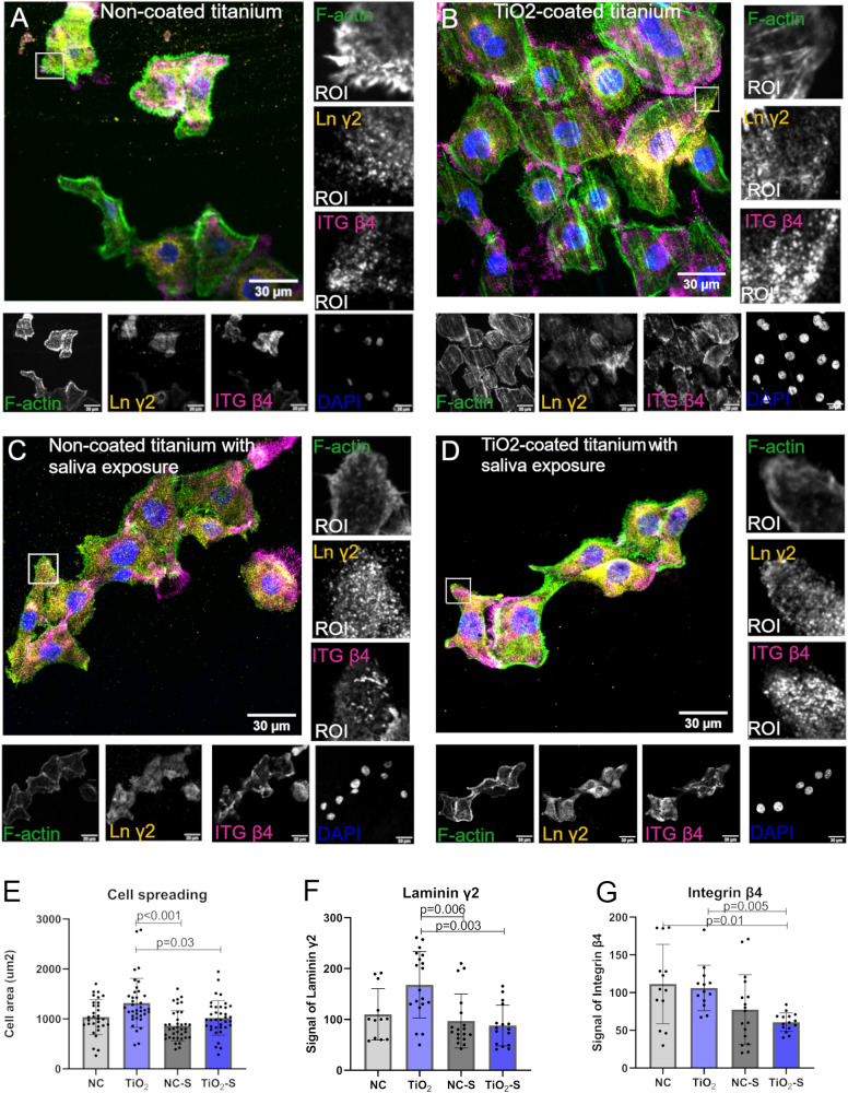
Fig. 4.
Saliva exposure reduces cell spreading and adhesion protein expression on TiO2-coated surface. A–D Representative confocal microscope images from the bottom plane of gingival keratinocytes stained with laminin γ2, integrin β4, dapi as nucleus and F-actin. Quantifications of (E) cell areas and signals of (F) Laminin γ2 and (G) Integrin β4. ROI region of interest. Mean ± SD, each data point represents a technical measurement, Kruskal Wallis test

