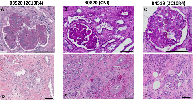
Fig. 3. Three of five xenograft recipients with histologic evidence of rejection at time of necropsy.
Top panels are periodic acid-Schiff staining of xenograft kidney biopsy sample derived from recipients B3520 (A), B0820 (B), and B4519 (C) at the time of necropsy; Hematoxylin and eosin (H&E) staining of B3520 (D), B0820 (E), and B4519 (F) at time of necropsy. Multiple cuts of each sample were obtained and evaluated with similar findings. The black bar at the bottom right of each panel corresponds to 100 μm.

