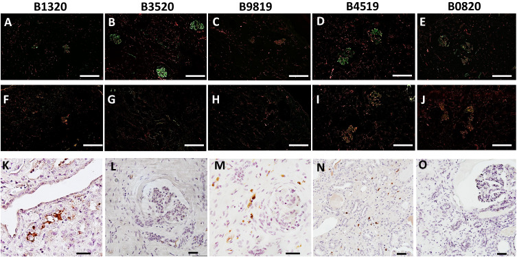
Fig. 4. Xenograft loss in remaining animals associated with adenovirus infection in the absence of antibody binding in xenograft.
Immunofluorescence staining of IgM (upper panels, A–E) and IgG (middle panels, F–J) of 10GE xenograft biopsies obtained from recipients at time of necropsy, double-stained with CD31 and performed in duplicate to confirm findings. White bar at the bottom right of each (A–J) corresponds to 200 µm. Immunohistochemistry staining for human adenovirus (lower panels, K–O); staining performed in duplicate to confirm findings. Black bar at bottom right of each (K–O) corresponds to 40 µm.

