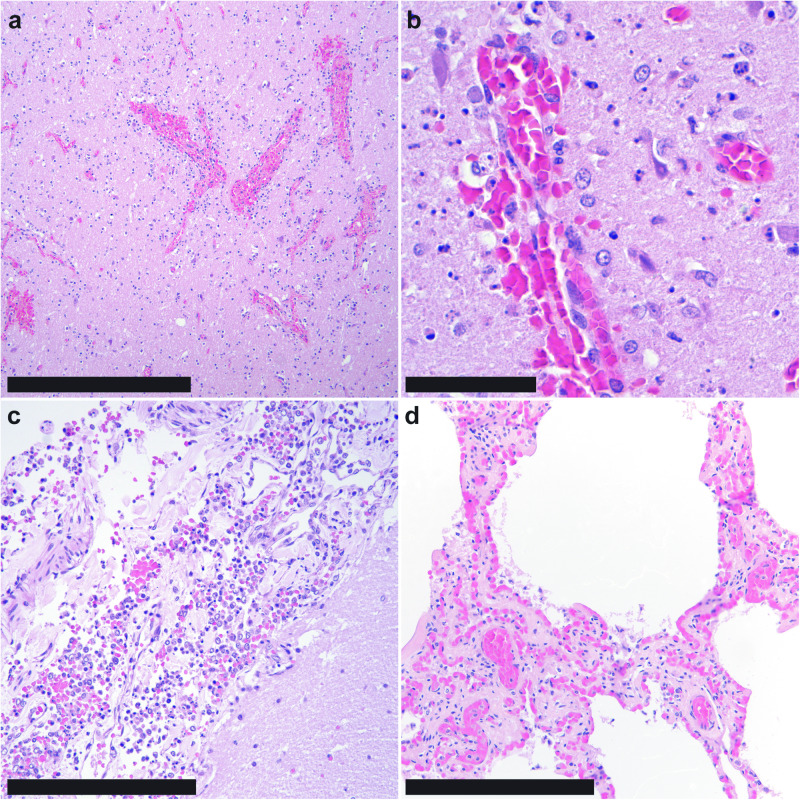
Fig. 1. Microscopic features of influenza A(H5N1) virus infection in a common bottlenose dolphin (Tursiops truncatus).
a In the brain, the cerebral neuroparenchyma is hypercellular with areas of congestion, hemorrhage, and loose, mononuclear perivascular cuffs; bar = 500 µm. b Within the affected neuropil, there is frequent necrosis of neurons and glial cells, as characterized by cytoplasmic eosinophilia and nuclear fragmentation; bar = 50 µm. c The leptomeninges are expanded by mixed mononuclear cells and hemorrhage; bar = 250 µm. d The pulmonary parenchyma was largely unremarkable, changes consistent with mild postmortem autolysis; bar = 250 µm. Hematoxylin and eosin, all panels.

