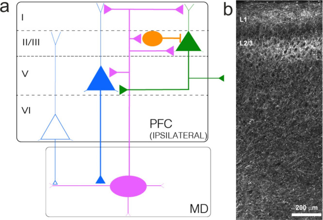
Figure 1.

Prefronto-thalamic projections. (a) Schematic overview of MD–PFC inputs. MD neurons predominantly project to superficial layers, and, to a lesser extent, to deep layers of the PFC. MD projections to layer I mostly target distal dendrites of PNs located in other layers. In layer II/III, MD neurons project to both PNs and interneurons. Interneurons excited by MD axons form a feedforward inhibitory microcircuit. Layer II/III PNs project to neurons in other layers or the contralateral hemisphere. MD neurons also project to layer V pyramidal neurons. Layers V and VI are the main output-generating layers in the PFC and project it to the MD. Inputs from layers V and VI differ in their morpho-functional properties. Colors of triangles represent subpopulations of pyramidal neurons residing in different layers. Round PFC neurons represent PV IN. Oval-shaped MD neurons represent a thalamic excitatory neuron. (b) Projection of MD axons to the PFC. Micrograph illustrating infection of AAV-ChR2-mCherry in the MD. mCherry-positive fibers can be detected in medial PFC. MD axons project both to superficial and deep layers. Note the dense accumulation of fibers in superficial and middle layers of the mPFC.
