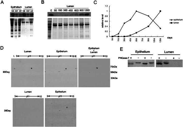
Fig. 10.
Comparison of GLB1L4 in the rat epididymal epithelium and lumen by Western blot analysis. A) Western blot analysis of GLB1L4 from rat epididymal epithelium and lumen protein extracts of the caput (ca), corpus (cor), and cauda (cd) regions. Protein (30 μg) was loaded in each lane. The same volume of protein was separated by electrophoresis and stained by Coomassie blue to show the equal loading. B) Western blot analysis of GLB1L4 from epididymal lumen protein during development. Protein (20 μg) was loaded in each lane. The same volume of protein was separated by electrophoresis and stained by Coomassie blue to show the equal loading. E, epididymis; D, days. C) Quantitative analysis of the relative protein expression levels in epididymal epithelium and lumen fluid at different ages in days (d). Protein extracts were pooled from three animals per group. D) Lumen (100 μg), epididymal epithelium (100 μg), and epithelium plus lumen (200 μg epithelium and 100 μg lumen) proteins were separated by 2D gel electrophoresis and detected using anti-GLB1L4 antisera. Molecular mass is indicated on the right (kDa), and pI values are shown on the top. E) The change in molecular mass of mature (90-day-old; left) and adolescent (30-day-old; right) rat GLB1L4 before (−) and after (+) deglycosylation by PNGase-F. Epithelium, epididymal epithelium protein; Lumen, lumen fluid protein.

