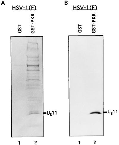
FIG. 3.
Autoradiogram and photograph of proteins from infected cell lysates that were bound to GST or GST-PKR, subjected to electrophoretic separation and autoradiography, and reacted with the anti-US11 antibody. Equivalent quantities of GST or GST-PKR bound to beads were reacted overnight with the radiolabeled, RNase-treated, and sonicated lysates from 2 × 10⁶ HeLa cells infected with HSV-1(F) as described in Materials and Methods. The beads were then washed seven times with PBS containing 1% NP-40 and 1% DOC, denatured in disruption buffer, boiled, subjected to electrophoresis in a denaturing 12.5% polyacrylamide gel, electrically transferred to a nitrocellulose sheet, reacted with the anti-US11 antibody, and subjected to autoradiography. The position of the US11 protein band is indicated. (A) Autoradiographic image of the electrophoretically separated proteins bound to beads; (B) photograph of the immunoblot.

