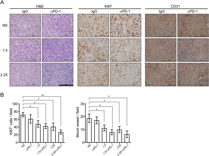
Fig. 6.
Combining anlotinib and PD-1 blockade inhibiting tumor proliferation and angiogenesis. a Representative images of H&E, Ki67, and CD31 staining in LLC-derived xenograft tumors. Images all were obtained at × 400 magnification; scale bar = 100 μm. b Quantification of CD31-positive vessels and Ki67-positive cells in the primary tumors. Results are the means ± SEM, n = 5, one-way ANOVA. *p < 0.05, **p < 0.01

