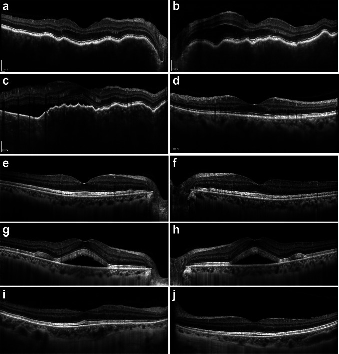
Fig. 1.
Representative enhanced depth imaging optical coherence tomography images of patients with ellipsoid zone thickening/serous retinal detachment or chorioretinitis after immune checkpoint inhibitor therapy. a, b A 58-year-old female (Patient 2) presented with bilateral chorioretinitis/papillitis 47 days after pembrolizumab therapy. c, d, A 54-year-old female (Patient 3) presented with chorioretinitis/papillitis 51 days after pembrolizumab therapy (c). Undulating retinal pigment epithelium, subretinal fluid, and thickened was resolved after posterior subtenon triamcinolone injection and systemic steroid treatment (d). e–h A 54-year-old man (Patient 30) presented with bilateral ellipsoid zone thickening (e, f) 13 days after atezolizumab/cobimetinib therapy. Serous retinal detachment developed after five cycles of atezolizumab/cobimetinib therapy (g, h). i, j A 66-year-old woman (Patient 15) presented with bilateral ellipsoid zone thickening and serous retinal detachment 4 days after pembrolizumab/trametinib therapy. No abnormality was detected during pembrolizumab monotherapy in this patient

