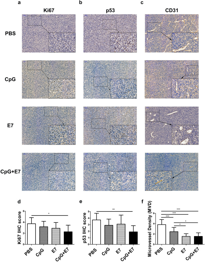
Fig. 5.
Effect of the vaccine on cellular proliferation, apoptosis and vessel density in tumors. Fewer a, d Ki67+ and b, e p53+ cells were quantified in mice treated with the vaccine compared to the control mice. c, f The expression of CD31 was significantly decreased in the vaccine group compared to the control group. Representative images of a Ki67, b p53 and c CD31 staining are shown. Images are shown at 100× magnification. Scale bars = 100 μm. Insert images are at 400× magnification and scale bars = 50 μm. The data are depicted as the mean ± SD. The significance of the data was evaluated by one-way ANOVA followed by Tukey’s multiple comparison test (*p < 0.05, **p < 0.01, ***p < 0.001)

