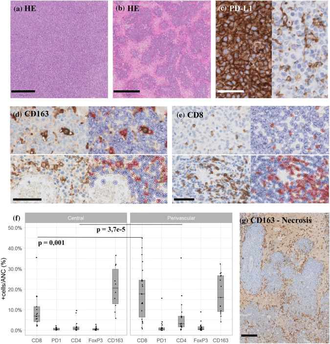
Fig. 1.
Immunohistochemistry analysis. a Hematoxylin and eosin (HE) staining of a non-necrotizing case, scale bar 500 µm; b HE staining of a necrotizing PCNSL, scale bar 500 µm; c PD-L1 IHC illustrating staining of both neoplastic and phagocytic cells (left) versus staining of only phagocytes (tumor negative; right), scale bar 50 µm; d CD163 IHC (left) and Qupath overlay of positive/red and negative/blue cells (right) in both central tumor bulk (top panels) and perivascular (bottom panels), scale bar 100 µm; e CD8 IHC (left) and Qupath overlay of positive/red and negative/blue cells (right) in both central tumor bulk (top) and perivascular (bottom), scale bar 100 µm; f boxplots of average positive % ANC of IHC markers; g CD163 IHC of a necrotizing lymphoma biopsy with lining of the tumor by CD163+ cells

