Fig. 1.
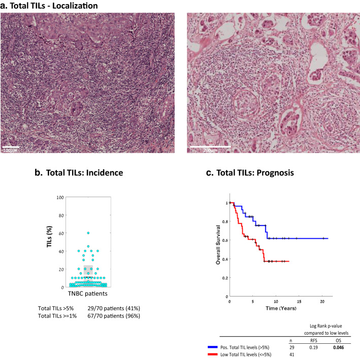
The presence of TILs in TNBC patient tumors is significantly associated with improved survival. The presence of TILs was determined in the 70-patient TNBC cohort used in our study. a Representative images of TIL localization in two patient tumors, demonstrated by H&E staining. b Percentage of TILs in each patient tumor (=dot), out of the total cellular tumor mass. Black line, Mean; Light gray box, Standard deviation; Dark grey box, SEM at 95% confidence interval. c OS Kaplan-Meier plot comparing patients with “Positive” (> 5%) vs. “Low” (≤ 5%) TIL levels. The corresponding RFS Kaplan-Meier plot is provided in Supplementary Figure 1a. +, Censored. p values of OS and RFS analyses are provided in the respective Figures

