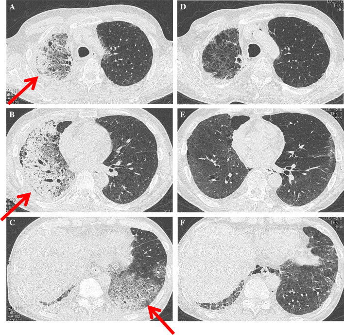
Fig. 1.
a–c Chest CT scan on admission (after 3 weeks of initial pembrolizumab administration) showing non-segmentally extended consolidations and GGOs with a predominant subpleural distribution in the whole lobes of the right lung and the lower lobe of the left lung (arrows). Traction bronchiectasis and bronchiolectasis with volume loss were also demonstrated. d–f Chest CT scan after triple combination therapy showing the marked amelioration of consolidations and GGOs in the bilateral lungs

