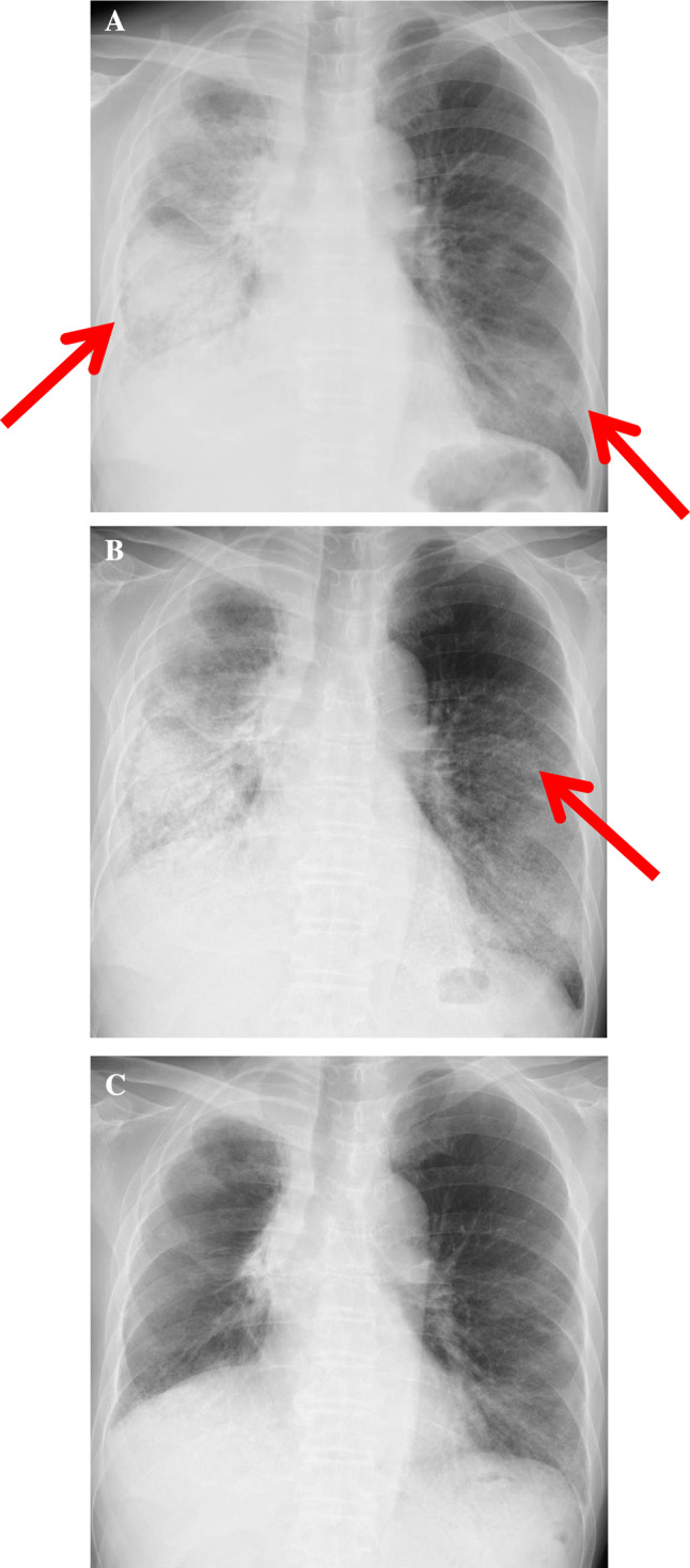
Fig. 2.

a Chest radiography on admission (after 3 weeks of the initial pembrolizumab administration) showing extensive consolidations and GGOs in the right whole pulmonary fields and the left lower pulmonary field (arrows). b Chest radiography in the ICU (after methylprednisolone pulse therapy) showing the expansion of GGOs in the left middle pulmonary field (arrow). c Chest radiography after triple combination therapy showing the improvement of the pulmonary opacities in the bilateral lungs
