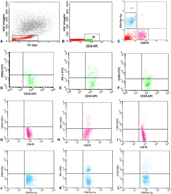
Fig. 1.
Flow cytometric detection of lymphocytes and their expression of TIM-3, PD-1, and CCR5. a Forward and side scatter histogram was used to define the lymphocyte population. b, c The expression of CD19, CD8 and CD4 was assessed on the lymphocytes, and then, the CD19+, CD8+, and CD4+ lymphocyte subsets were gated for further analysis of TIM-3, PD-1, and CCR5. d–f The expression of Tim-3, PD-1, and CCR5 on CD19. g–i The expression of Tim-3, PD-1, and CCR5 on CD8. j–l The expression of Tim-3, PD-1, and CCR5 on CD4

