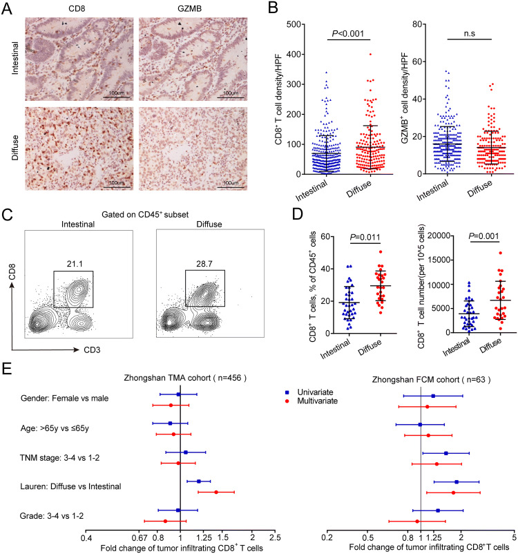
Fig. 1.
Distribution of tumor-infiltrating CD8+ T cells by Lauren classification. a Representative immunohistochemistry images of CD8 and GZMB staining in gastric cancer tissues with different Lauren subtypes (200× magnification). b Density of CD8+ and GZMB+ T cell infiltration in intestinal type (n = 286) and diffuse type (n = 170) gastric cancer patients from Zhongshan TMA cohort. c Representative images from flow cytometry showing CD8+ T cells proportions in intestinal type and diffuse type gastric cancer. d Proportions among all CD45+ leukocytes and absolute numbers of tumor-infiltrating CD8+ T cells in intestinal type (n = 36) and diffuse type (n = 27) gastric cancer patients from Zhongshan FCM cohort. e Univariate and multivariate associations between tumor-infiltrating CD8+ T cells and clinicopathological variables in Zhongshan TMA cohort and FCM cohort. Data are represented as mean ± SD, and P values are calculated using unpaired two-tailed t test. GZMB granzyme B, TMA tissue microarray, FCM flow cytometry

