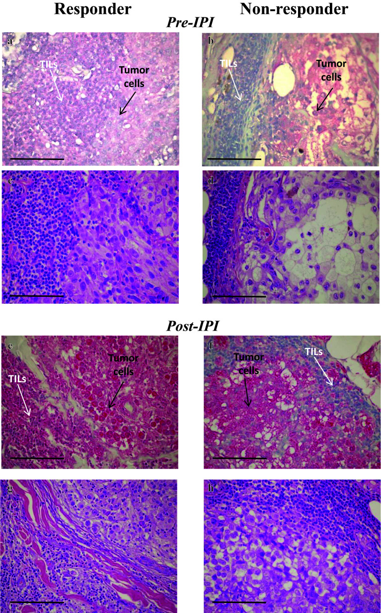
Fig. 3.
Representative immunohistochemical (IHC) stainings of CTLA-4 expressing TILs in melanoma tissues from patients responding (R) and non-responding (NR) to IPI therapy. CTLA-4 IHC staining was performed, with the antibody 14D3, on formalin-fixed, paraffin-embedded tissue sections from melanoma patients before (pre-IPI) and after (post-IPI) treatment with ipilimumab (IPI). Antibody reactivity was detected with AP-conjugated secondary antibody and Fast Red chromogen (red staining), as described in the section “Materials and methods”. Slides were counterstained with hematoxylin before microscopical evaluation. a and e Melanoma tissues from two R patients showing CTLA-4 positively stained TILs (white arrows) distributed along the invasive margin at the left of the tumor cells. b and f Melanoma tissues from two NR patients showing CTLA-4 negative TILs (white arrows) distributed along the invasive margin at the left (b) and at the right (f) of the tumor cells. Tumor cells show a moderate CTLA-4 positivity (a, b) and a strong CTLA-4 positivity (e, f). H&E, haematoxylin and eosin (c, d, g, h). Original magnification: 60×. Scale bar, 100 µm. TIL tumor infiltrating lymphocytes

