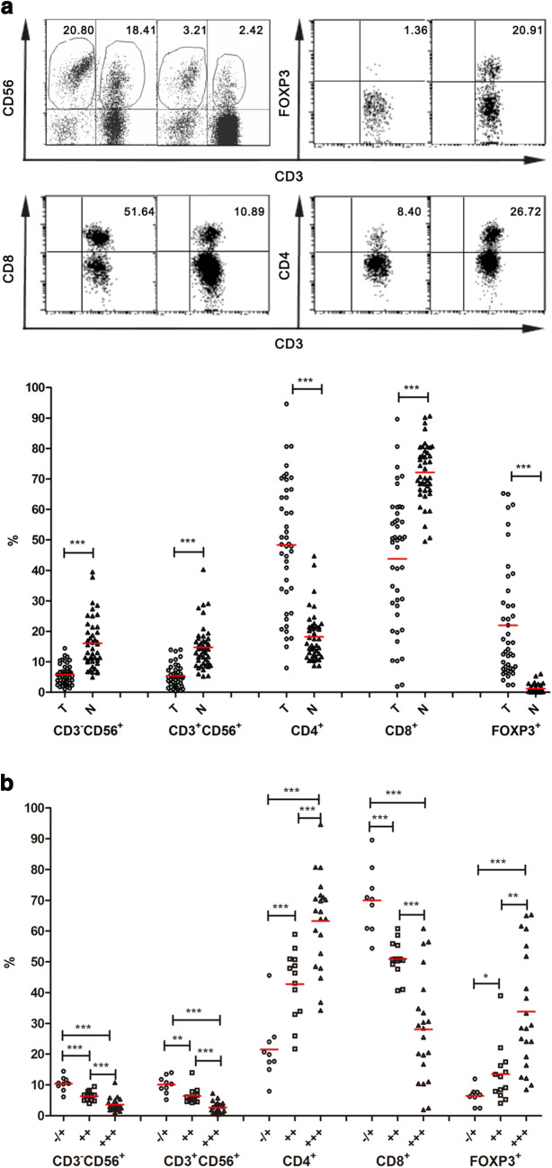
Fig. 4.
Comparative analyses of the TIL repertoires in HCC tissues with different levels of CD147 expressions. a Infiltrating lymphocyte compositions in tumor tissues and adjacent matching normal tissues obtained from patients with HCC, including the percentages of NT (CD3+CD56+) and NK (CD3−CD56+), CD4+, CD8+ and FOXP3+ subsets in CD3+ lymphocytes were analyzed using flow cytometry. A representative profile of scatter plots was shown (upper). b Based on the levels of CD147 expression (∓, ++, +++) in tumorous tissues of HCC, TILs were divided into three groups. A comparative analysis was performed between those three groups for each lymphocyte subsets mentioned above. *p < 0.05, **p < 0.01, ***p < 0.001

