Fig. 3.

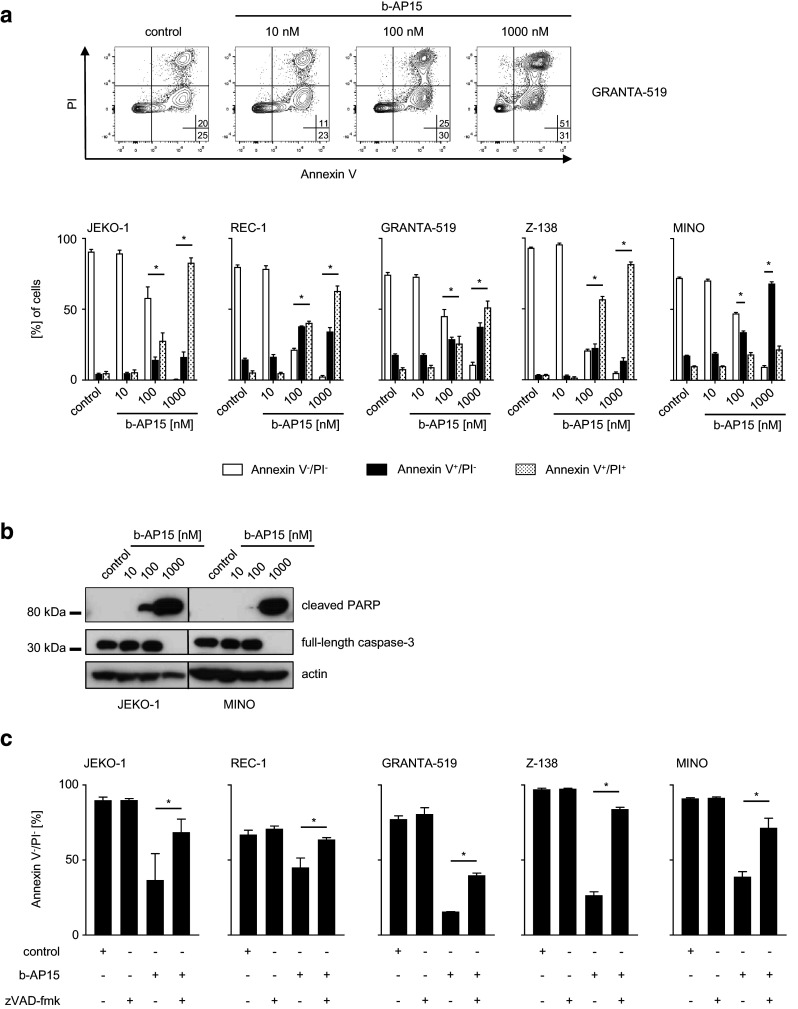
b-AP15 induces caspase-dependent apoptosis in MCL cells. a, b MCL cells were exposed to the indicated concentrations of b-AP15 or DMSO for 24 h. The percentage of live (Annexin V−/PI−), early (Annexin V+/PI−) and late apoptotic/necrotic (Annexin V+/PI+) MCL cells was determined by FACS with Annexin V/PI staining. Upper panel represents exemplary dot plot data obtained with the indicated MCL cell line; in the lower panel, combined data of three independent experiments with the indicated MCL cell lines is shown. Student’s t test was used to determine statistical significance of differences between control and treated cells (*p < 0.05). b Whole-cell lysates were subjected to western blot analyses using antibodies directed against full-length caspase-3 and cleaved PARP with actin serving as loading control. One representative result of at least three experiments with similar results is shown. c MCL cells were exposed to b-AP15 (500 nM) with or without zVAD-fmk (20 µM) or to zVAD-fmk or DMSO alone for 24 h. Data are presented as percentage of viable (Annexin V−/PI−) cells relative to control as determined by Annexin V/PI staining. Data represent means of three technical replicates. Bars represent SD; Student’s t test was used to determine statistical significance of differences between b-AP15 cells treated with or without additional exposure to zVAD-fmk (*p < 0.05)
