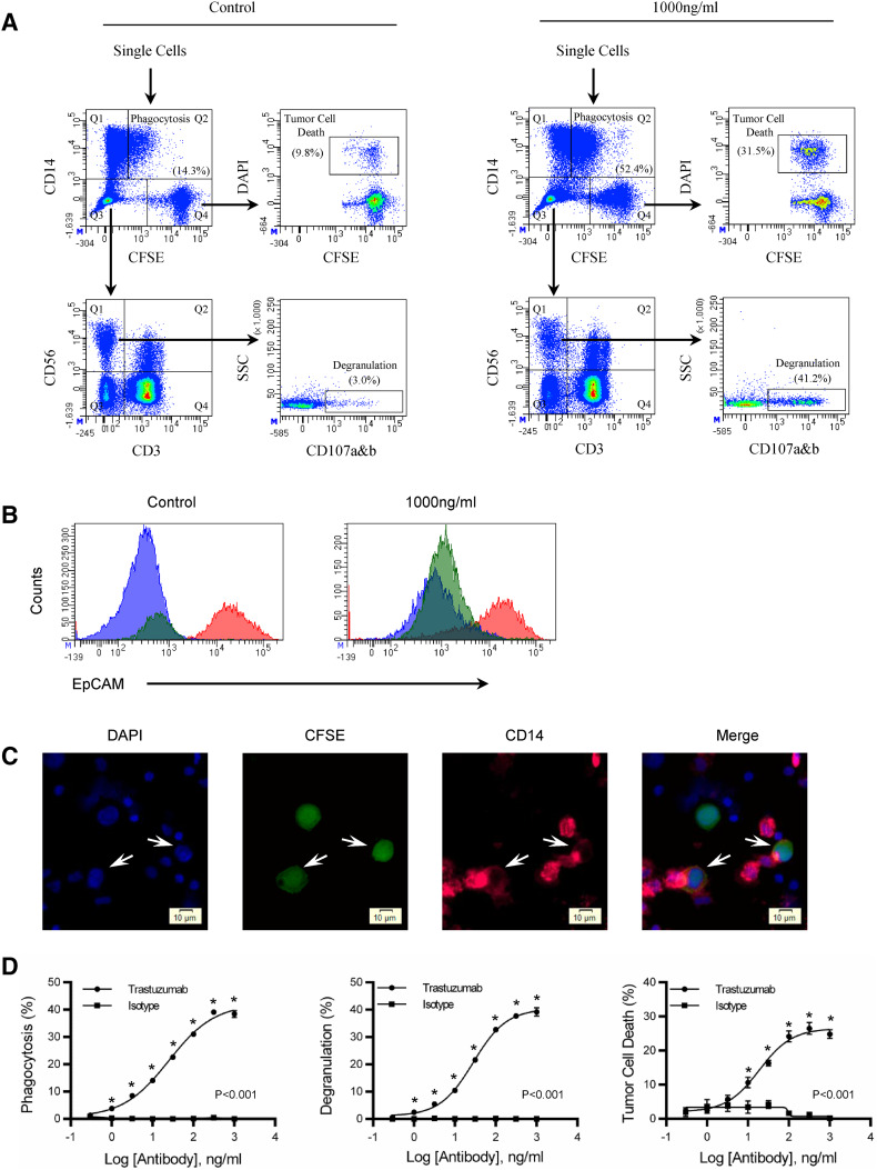
Fig. 1.
Simultaneous evaluation of antibody-dependent phagocytosis, NK cell degranulation, and tumor cell death. A 10:1 PBMC-to-tumor ratio was used to evaluate antibody-mediated effector function over a 4 h period. a See the “Results” section of this manuscript for a description of the gating strategy. Flow cytometry dot plots show control and 1000 ng/ml of trastuzumab. Values of phagocytosis, degranulation, and tumor cell death are in parenthesis. b To confirm phagocytosis, a second-tumor antigen EpCAM was used to determine if the monocytes internalized the tumor or not. Tumor cells (Q4) and monocyte (Q1 and Q2) were gated from the CD14 versus CFSE plot. Tumor cells are the red histogram, monocytes that have phagocytized tumors are the green histogram, and monocytes without tumor cells are the blue histograms. c Microscopy showing monocyte phagocytosis of tumor cells. Nuclei are stained with DAPI (Blue), HCC1419 (Green), and CD14 monocytes (Pink). Trastuzumab was used at 100 ng/ml and the images are from a trastuzumab co-culture of PBMC and HCC1419. d Titration curves of degranulation, tumor cell death, and phagocytosis comparing trastuzumab with isotype control. Background levels of phagocytosis, degranulation, and tumor cell death are subtracted from experimental values. All samples were performed in triplicate; means ± SEM and regression line are plotted on the graph. Two-way ANOVA with Sidak’s multiple comparisons test was used to determine significance. The asterisk represents significance between trastuzumab and isotype control. Results are representative of three independent experiments

