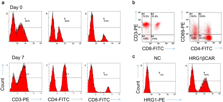
Fig. 2.
Expression of HRG1β-CAR in activated T lymphocytes. a PBMCs were isolated from healthy volunteers’ blood. After isolation, cells were labeled with anti-CD3-PE, anti-CD4-FITC, anti-CD8-FITC, and detected by flow cytometry. b CD3-positive T cells were obtained from PBMC by magnetic bead separation, 1 week after anti-CD3/CD28 activated culture, and were subjected to flow cytometry analysis. c Expression of HRG1β was detected by flow cytometry in activated T-lymphocytes transduced with HRG1β-CAR. Data are representative of PBMCs from five donors

