Fig. 5.
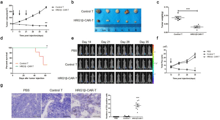
T-lymphocytes expressing HRG1β-CAR exert antitumor activity in vivo. a–c Nude mice (n = 10) were injected subcutaneously with SK-BR-3 cells in the right back to form xenograft tumors, followed by random grouping and weekly tail vein administration of control or HRG1β-CAR-T cells. Tumor volume was monitored and plotted (a). Mice were killed on day 42, and tumors were excised and weighed (b, c). d Xenograft tumors were established and mice were treated as described in (a–c, n = 6 for each group). The survival of mice was monitored and plotted. e–g Nude mice were inoculated with SK-BR-3 cells expressing luciferase (n = 3 for each group), followed by tail intravenous treatment with PBS, control T and HRG1β-CAR-T cells on day 14. Bioluminescent imaging was performed on indicated days (e), and region-of-interest (ROI) bioluminescence emission measurement was calculated for each group (f). Mice were killed on day 35, and tumors were excised, sectioned and subject to immunohistochemical staining for CD3 (g). The numbers of CD3-positive cells in 9 independent microscope fields were plotted. Magnification, ×400. Scale bar = 100 μm. *P < 0.05, **P < 0.01 (Student’s t test)

