Fig. 3.
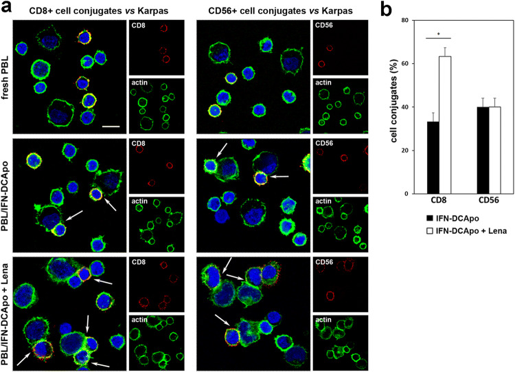
Effects of lenalidomide on conjugate formation. a CLSM examinations of lymphoma co-cultures (central optical sections) with freshly purified PBL or PBL cultured for 14 days with tumor cell-loaded IFN-DC in the absence or presence of lenalidomide. Cells were stained for CD8 or CD56 (red) and for actin detection (green). DAPI was used to stain nuclei (blue). Arrows indicate CD8 + or CD56 + cell conjugates. Scale bars 10 µm. Images are representative of three independent experiments. b Quantitative analysis of cell conjugates expressed as the median percentage of CD8 + or CD56 + cell conjugates from PBL/lymphoma co-cultures treated or not with lenalidomide (median ± SE of three independent experiments). Statistically significant differences were estimated by the Wilcoxon test (*p < 0.05)

