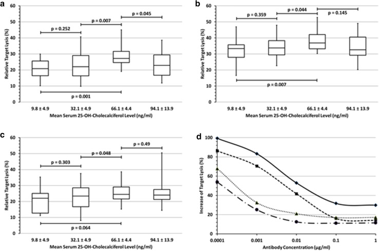
Fig. 3.
Effect of vitamin D3 levels on NK cell-mediated ADCC a–c summarize the ADCC-mediated lysis rates of all individuals at four actually achieved 25-OH-D3 serum levels at an E:T ratio of 2.5:1 and 0.1 µg/ml of rituximab-loaded DAUDI cells (a), obinutuzumab-loaded DAUDI cells (b) and trastuzumab-loaded ZR-75-1 breast cancer cells (c). 25-OH-D3 substitution restricted to the lower normal range (32.1 ng/ml) did not affect the NK cell-mediated ADCC. After continued substitution only rituximab- (p = 0.001) and obinutuzumab-mediated ADCC (p = 0.007) increased significantly at a 25-OH-D3 serum level of 66.1 ng/ml compared to base line, while the increase of trastuzumab-mediated ADCC was not significant (p = 0.064). Further 25-OH-D3 substitution to 94.1 ng/ml resulted in a weak, but significant decline of the rituximab-mediated ADCC (p = 0.045) compared to the maximum, while this decline was minimal for obinutuzumab (p = 0.145) and remained unchanged for trastuzumab (p = 0.49). d Relative 25-OH-D3 induced increase of target cell lysis at different antibody concentrations and E:T ratios after the substitution of 25-OH-D3 serum levels to 66.1 ng/ml. The curves show the decreasing impact of 25-OH-D3 with increasing antibody concentrations and higher E:T ratios and the smaller effect on obinutuzumab-ADCC compared to rituximab-ADCC. Signs and symbols: Check/closed line: rituximab (E:T = 2.5:1); quadrate/dashed line: rituximab (E:T = 5:1); triangle/dotted line: obinutuzumab (E:T = 2.5:1); circle/chain line: obinutuzumab (E:T = 5:1)

