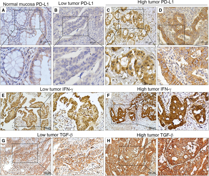
Fig. 1.
Representative PD-L1, IFN-γ and TGF-β immunohistochemical images from TMA patients with LARC. a PD-L1 expression in normal mucosa. b–d Low and high tumor PD-L1 expression in pre-neoCRT biopsies. e, f Low and high tumor IFN-γ expression in post-neoCRT tissues. g, h Low and high tumor TGF-β expression in post-neoCRT tissues

