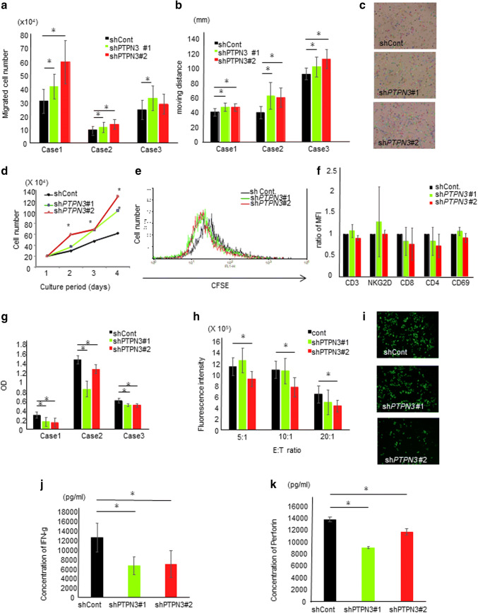
Fig. 2.
PTPN3 inhibition augments the functions of activated T lymphocytes from healthy volunteers in vitro. a The migration ability of lymphocytes of the day of the 7 day culture was analyzed by the chamber method. b The migration ability of lymphocytes of the day of the 7 day culture was analyzed by time-lapse imaging. The graph shows the moving distance of lymphocytes analyzed by the Image-Pro Analyzer software program. c Representative pictures of time-lapse imaging. The tracking line shows the moving distance. Original magnification ×20. d The proliferation of lymphocytes was estimated. Cell numbers at the indicated days were counted using a light microscope. e 5 days after activation, lymphocytes were labeled by CFSE, and after an additional 2 days of culture, the immunofluorescence intensity of CFSE in lymphocytes was analyzed by FACS. f The surface antigen expression of lymphocytes was analyzed by FACS. g The cytotoxicity of lymphocytes after cancer cells and lymphocytes were co-cultured was analyzed. After WST reagent solution had been added to the well, the absorbance of viable cancer cells was detected by subtracting the absorbance of lymphocytes alone from that of co-culture. OD shows the calculated absorbance of viable cancer cells. h The cytotoxicity of lymphocytes was estimated by time-lapse imaging. After co-culture of lymphocytes and calcein-labeled SCC cells, the fluorescent intensity of viable cancer cells was measured by the Image-Pro Analyzer software program. i Representative pictures of time-lapse imaging of viable cancer cells. Original magnification ×20. The IFN-γ (j) and perforin (k) secretion capacity of lymphocytes was measured by an ELISA. The lymphocytes used in each experiment of a–g were obtained from three different healthy volunteers. Similar results were obtained in two different healthy volunteers (h–k). Data are presented as the mean ± SD. *p < 0.05

