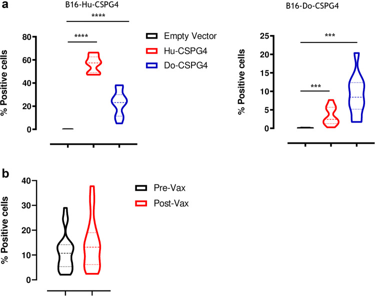
Fig. 3.
Anti-CSPG4 vaccine-induced antibody response. a Anesthetized C57BL/6 mice were vaccinated as previously described in [94] and sera collected 2 weeks after vaccination were tested for their ability to stain murine B16 melanoma cells stably transfected with either the human (left panel) or canine (right panel) CSPG4 antigen. Results are expressed as percentage (%) of CSPG4 positive cells. Student’s t test ***P < 0.0006; ****P < 0.0001. b Canine MM patients were vaccinated with the Hu-CSPG4 DNA plasmid, as previously described in [39, 40], and sera collected before the first immunization (Pre-Vax) and after the fourth vaccination (Post-Vax) were selected for further analysis. Sera were tested for their ability to stain the canine CSPG4 antigen on the canine CSPG4+ MM cell line (CMM-12). The IgA-specific binding was revealed using a goat anti-dog IgA secondary antibody. Results are expressed as percentage (%) of CSPG4 positive cells. Flow cytometry was performed using a FACS Verse (BD Biosciences) and the results were analyzed using BDFacs Suite software

