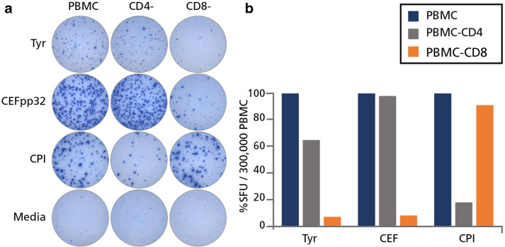
Fig. 3.
Tyrosinase-triggered IFN-γ spots are produced primarily by CD8 cells. PBMC from HD #36 were tested “unfractionated”, or after depletion of CD4 (CD4−) or CD8 (CD8−) cells via magnetic beads. These cells were then cultured either with tyrosinase (Tyr), CEF peptides (CEFpp32), CPI, or left unstimulated (media) and IFN-γ was detected after 24 h. a The original wells are shown. b The percentage reduction in spot forming unit (SFU) counts is shown after CD4 or CD8 cell depletion, representing the response of unfractionated PBMC as 100%

